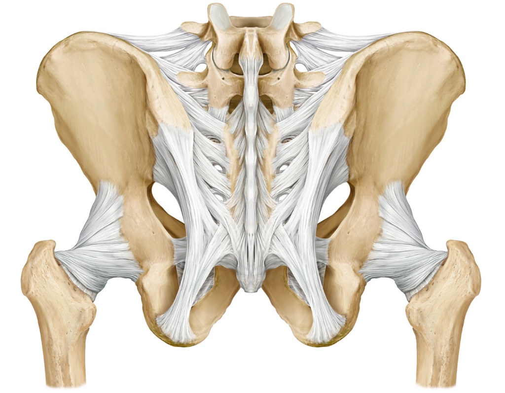
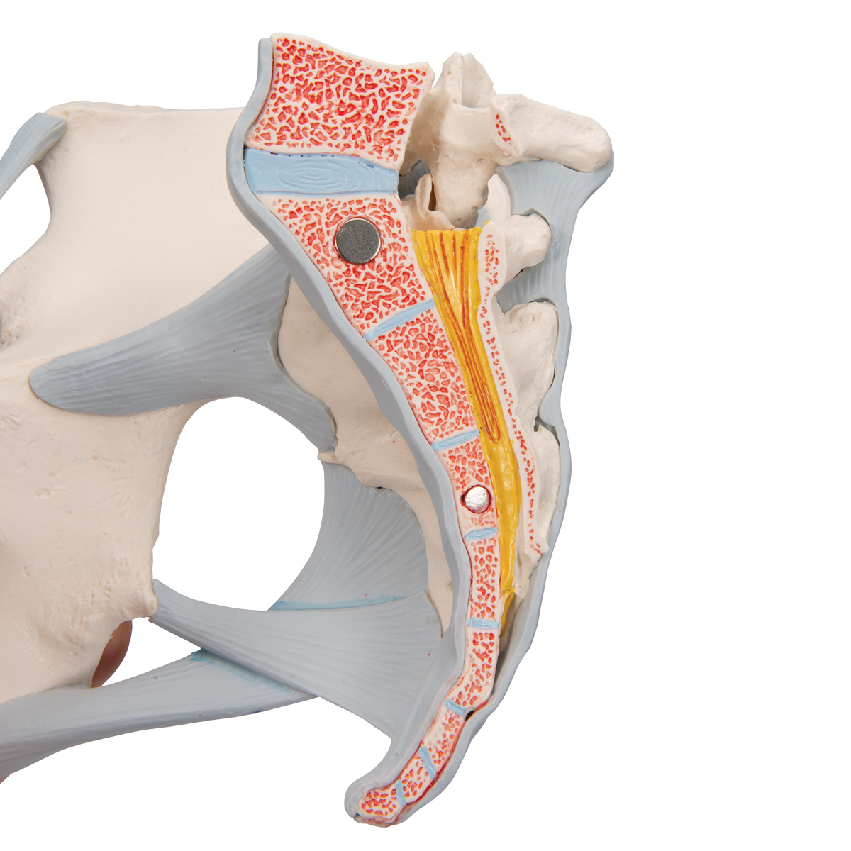
Pelvic Anatomy Ligaments / The named ligaments of the pelvis mostly arise from the sacrum and attach to varying segments of the pelvic bone.. The broad ligament overlies the structures and connective tissue immediately adjacent to the uterus. 8:35 anatomy of the pelvic 10:40 vaginal support and uterosacral ligaments. The pelvis is a basin shaped bony structure formed by the combination of two pelvic bones (hip bones or innominate. Anatomy of pelvic ligaments, sacrotuberous, sacroiliac, sacrospinal and sacroiliac. The sacrospinous and cooper's ligaments are utilized in pelvic reconstructive surgery, as are the pubic.
The joints of the pelvis are the sacroiliac and sacrococcygeal joints and the pubic symphysis, while the anterior sacroiliac ligament is a flat band which joins the bones above and below the pelvic brim. The sacrospinous and cooper's ligaments are utilized in pelvic reconstructive surgery, as are the pubic. Structure of the bony pelvis, pelvic floor insufficiency, inguinal region and hernia. Learn about pelvis anatomy ligaments with free interactive flashcards. Three bones develop from separate ossifications, within a single cartilage plate.
Ligaments - Posterior Pelvis | Donny Perry | Flickr from c2.staticflickr.com The pelvis (plural pelves or pelvises) is either the lower part of the trunk of the human body between the abdomen and the thighs (sometimes also called pelvic region of the trunk) or the skeleton embedded in it (sometimes also called bony pelvis, or pelvic skeleton). Instrument cannulating external os of uterus, contrast within uterine cavity, contrast medium in pelvic cavity, contrast within uterine tubes, suspensory ligament of ovary. The broad ligament overlies the structures and connective tissue immediately adjacent to the uterus. Surgical pelvic anatomy in gynecologic oncology. Functional anatomy of the anterior cruciate ligament. 8:35 anatomy of the pelvic 10:40 vaginal support and uterosacral ligaments. Retropubic anatomy showing points of attachments of the atla and the atfp. Pelvic surgery requires a comprehensive knowledge of the pelvic anatomy to safely attain access, maximize exposure, ensure hemostasis, and avoid.
With inks to related posts.
Functional anatomy of the anterior cruciate ligament. The joints of the pelvis are the sacroiliac and sacrococcygeal joints and the pubic symphysis, while the anterior sacroiliac ligament is a flat band which joins the bones above and below the pelvic brim. 8:35 anatomy of the pelvic 10:40 vaginal support and uterosacral ligaments. Surgical pelvic anatomy in gynecologic oncology. The pelvic girdle consists of two symmetrical halves. Amis, a and g dawkins. Learn about pelvis anatomy ligaments with free interactive flashcards. Read more.it is secured by strong ligaments. Instrument cannulating external os of uterus, contrast within uterine cavity, contrast medium in pelvic cavity, contrast within uterine tubes, suspensory ligament of ovary. During pregnancy, the ligaments between the symphysis and the. The pelvis (plural pelves or pelvises) is either the lower part of the trunk of the human body between the abdomen and the thighs (sometimes also called pelvic region of the trunk) or the skeleton embedded in it (sometimes also called bony pelvis, or pelvic skeleton). There are two major groups of ligaments that provide nearly all the structure of the. Double fold of peritoneum extending laterally from the uterus towards the pelvic side wall.
Drawing of the pelvis indicating the main ligaments of the pelvis. The pelvic girdle consists of two symmetrical halves. Functional anatomy of the anterior cruciate ligament. Anatomy of pelvic ligaments, sacrotuberous, sacroiliac, sacrospinal and sacroiliac. Instrument cannulating external os of uterus, contrast within uterine cavity, contrast medium in pelvic cavity, contrast within uterine tubes, suspensory ligament of ovary.
Pelvic Anatomy Ligaments - Health, Medicine and Anatomy ... from i.pinimg.com • muscles and ligaments form a pelvic floor. 8:10 pelvic sidewall anatomy and retroperitoneal spaces. ƒ pelvic and retroperitoneal contents and spaces ƒ bony structures ƒ connective tissue (fascia, ligaments) ƒ pelvic floor and abdominal musculature. ƒ describe functional anatomy and relevant. With inks to related posts. The pelvis (plural pelves or pelvises) is either the lower part of the trunk of the human body between the abdomen and the thighs (sometimes also called pelvic region of the trunk) or the skeleton embedded in it (sometimes also called bony pelvis, or pelvic skeleton). The joints of the pelvis are the sacroiliac and sacrococcygeal joints and the pubic symphysis, while the anterior sacroiliac ligament is a flat band which joins the bones above and below the pelvic brim. Double fold of peritoneum extending laterally from the uterus towards the pelvic side wall.
Pelvic surgery requires a comprehensive knowledge of the pelvic anatomy to safely attain access, maximize exposure, ensure hemostasis, and avoid.
Choose from 500 different sets of flashcards about pelvis anatomy ligaments on quizlet. • muscles and ligaments form a pelvic floor. The broad ligament overlies the structures and connective tissue immediately adjacent to the uterus. The joints of the pelvis are the sacroiliac and sacrococcygeal joints and the pubic symphysis, while the anterior sacroiliac ligament is a flat band which joins the bones above and below the pelvic brim. Differences between the male pelvis and the female pelvis. Read more.it is secured by strong ligaments. • pelvis begins at the iliac crests and ends at the symphysis pubis. Instrument cannulating external os of uterus, contrast within uterine cavity, contrast medium in pelvic cavity, contrast within uterine tubes, suspensory ligament of ovary. Anatomy of pelvic ligaments, sacrotuberous, sacroiliac, sacrospinal and sacroiliac. Published on 09/03/2015 by admin. The named ligaments of the pelvis mostly arise from the sacrum and attach to varying segments of the pelvic bone. ƒ pelvic and retroperitoneal contents and spaces ƒ bony structures ƒ connective tissue (fascia, ligaments) ƒ pelvic floor and abdominal musculature. ƒ describe functional anatomy and relevant.
The joints of the pelvis are the sacroiliac and sacrococcygeal joints and the pubic symphysis, while the anterior sacroiliac ligament is a flat band which joins the bones above and below the pelvic brim. Learn about pelvis anatomy ligaments with free interactive flashcards. Published on 09/03/2015 by admin. An overview of uterine anatomy, including structure, relations, ligaments, blood supply and innervation. The pelvic girdle consists of two symmetrical halves.
Anatomical Teaching Models - Plastic Human Pelvic Models ... from www.a3bs.com ƒ describe functional anatomy and relevant. The pelvic girdle consists of two symmetrical halves. Functional anatomy of the anterior cruciate ligament. Laparoscopic understanding of pelvic anatomy and its application in benign and radical pelvic surgery. During pregnancy, the ligaments between the symphysis and the. The named ligaments of the pelvis mostly arise from the sacrum and attach to varying segments of the pelvic bone. • pelvis begins at the iliac crests and ends at the symphysis pubis. Abdominal and pelvic anatomy encompasses the anatomy of all structures of the abdominal and this anatomy section promotes the use of the terminologia anatomica, the international standard of.
There are many organs that sit in the pelvis, including much of the urinary system, and lots of the male or female reproductive systems.
Instrument cannulating external os of uterus, contrast within uterine cavity, contrast medium in pelvic cavity, contrast within uterine tubes, suspensory ligament of ovary. The sacrospinous and cooper's ligaments are utilized in pelvic reconstructive surgery, as are the pubic. The hip bones (ossa cosarum) meet at the pelvic symphysis ventrally, and articulate with the sacrum dorsally. ƒ describe functional anatomy and relevant. Introduction to pelvic anatomy 1. • muscles and ligaments form a pelvic floor. 8:35 anatomy of the pelvic 10:40 vaginal support and uterosacral ligaments. Various pelvic ligaments help support the uterus and other pelvic organs. Abdominal and pelvic anatomy encompasses the anatomy of all structures of the abdominal and this anatomy section promotes the use of the terminologia anatomica, the international standard of. Differences between the male pelvis and the female pelvis. During pregnancy, the ligaments between the symphysis and the. Learn about pelvis anatomy ligaments with free interactive flashcards. Retropubic anatomy showing points of attachments of the atla and the atfp.
Double fold of peritoneum extending laterally from the uterus towards the pelvic side wall pelvic anatomy. Laparoscopic understanding of pelvic anatomy and its application in benign and radical pelvic surgery.


0 Komentar